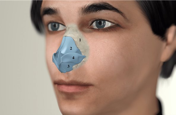
Structure of the nose
A nose is composed of inner and outer nasal structures.

Inner nasal cavity
Nasal septum
The inner nasal cavity is divided into the left and right nasal cavity by a cartilaginous-bony wall known as the nasal septum (3). There is cartilage in the anterior part of the nasal septum, which reduces in size with advancing age and turns into a bone, and there is a bony section in the posterior part, which extends to the nasopharynx.
The nasal septum is only a few millimetres wide at its widest point, however, differences in the rate of growth of the cartilaginous and bony parts can cause the nasal septum to shift to the left or to the right and lead to the formation of bony variations, such as a spur or a groin. A spur can cause extremely severe headaches in some patients.
The nasal septum is covered with mucous membrane, which exudes mucus that moistens the air we breathe and protects us from bacteria that would otherwise penetrate deeper into the lungs.
Nasal conchae
There are three elongated structures to the right and left of the main cavity, which are called nasal conchae. These consist of a small, elongated bone in the middle, embedded in a vascular corpus cavernosum and mucosa. The excretory ducts of the paranasal sinuses are located between the inferior and middle conchae.
The nasal conchae, along with the nasal septum, are responsible for ensuring that the airflow is linear and that the air is warmed and moistened. If the nasal septum is crooked on one side and/or the conchae are enlarged, less air gets through the nose. In the case of pronounced nasal breathing obstruction, a nasal septum straightening accompanied by nasal conchae reduction is recommended.
The techniques implemented in our practice are aimed at maximum tissue preservation. These procedures are usually tamponade-free.
If too much of the nasal conchae has been removed in a surgery, the mucous membranes in the nose often become too dry and it is observed that patients also complain of pronounced nasal breathing obstruction despite having enough space.
The outer nose shape
The shape of the outer nose depends on several factors:
1. The thickness of the skin and the underlying soft tissue
It is unfortunately not possible to achieve a petite nasal tip in patients with very thick, oily skin, because despite all the surgical measures taken on the underlying cartilaginous framework, an additional volume remains in the area of the tip.
The fat layer can only be partially removed, as extreme scarring may occur very quickly in this area. A cortisone injection should never be administered in non-pre-operated noses due to the potential for severe side effects such as scarring and irregularities.
2. The bony structure that forms the upper part of the nose
The bony part of the nose (1) consists of the two so-called nasal bones which form the bony nose in the continuation of the upper jaw. A nose can grow crooked, appear very flat, form a saddle nose or a pronounced hump due to an injury in adolescence. It may also be the result of a family predisposition. The most common condition responsible for an aesthetically unflattering nose is a nasal hump, which is usually made up of an excess layer of bone at the top and cartilage underneath.
3. The cartilaginous structure that defines the appearance of the lower half of the nose along with the skin
The cartilaginous shape of the nose consists of the interaction of five different cartilages: the nasal septum (3) in the middle, two lateral cartilages (2) on top of it like a roof, and two femoral cartilages (4) (outer and inner), which together with the skin give shape to the nasal wings and the nasal bridge.
When the nose has been affected by an injury, changes in the shape of the cartilage develop frequently as well. Particularly excessive growth of the nasal septum can also cause the entire nasal structure to elongate or the outer nose to become crooked. Strongly pronounced femoral cartilages can cause the so-called "bulbous nose".
